PSAP
PSAP,全稱為前鞘脂激活蛋白原,常被簡稱為prosaposin,也有SAP前體這樣的別名。它是體內脂質代謝的重要調節因子前體,可分解為四個功能片段,幫助細胞內溶酶體處理鞘脂類物質的降解與循環,維持細胞正常的脂質平衡狀態。除此之外,這種蛋白在神經系統中含量較高,具有明顯的神經保護作用,能支持神經細胞的存活與分化,減少有害物質對神經細胞的損傷。
若PSAP相關功能出現異常,可能導致鞘脂類物質在體內堆積,引發代謝性疾病,這類疾病對神經系統影響尤其顯著,患者可能出現認知減退、運動障礙等癥狀。目前針對PSAP的研究多聚焦于神經保護領域,部分團隊嘗試用其活性片段開發治療神經退行性疾病的候選藥物,比如緩解阿爾茨海默病的神經損傷,但相關研究仍處于早期階段,尚未有成熟藥物應用于臨床。
熱銷產品
Recombinant Human Proactivator polypeptide (PSAP), partial (CSB-EP018836HU)
驗證數據
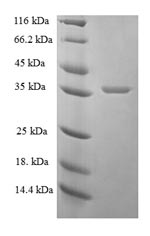
(Tris-Glycine gel) Discontinuous SDS-PAGE (reduced) with 5% enrichment gel and 15% separation gel.
PSAP Recombinant Monoclonal Antibody (CSB-RA264216A0HU)
驗證數據
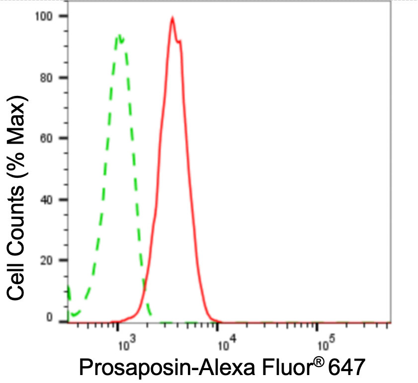
Flow cytometric analysis of prosaposin expression in HepG2 cells using prosaposin antibody. Green, isotype control; red, prosaposin.
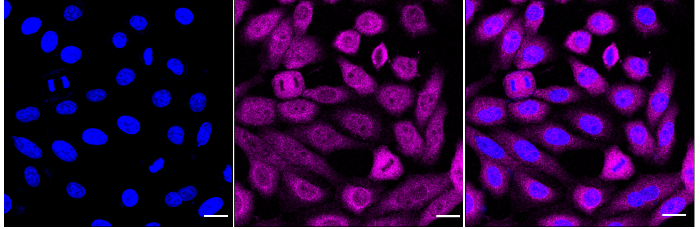
Immunocytochemical staining of HepG2 cells with prosaposin antibody. Nuclei were stained blue with DAPI; Prosaposin was stained magenta with Alexa Fluor? 647. Images were taken using Leica stellaris 5. Protein abundance based on laser Intensity and smart gain: Medium. Scale bar: 20 μm.
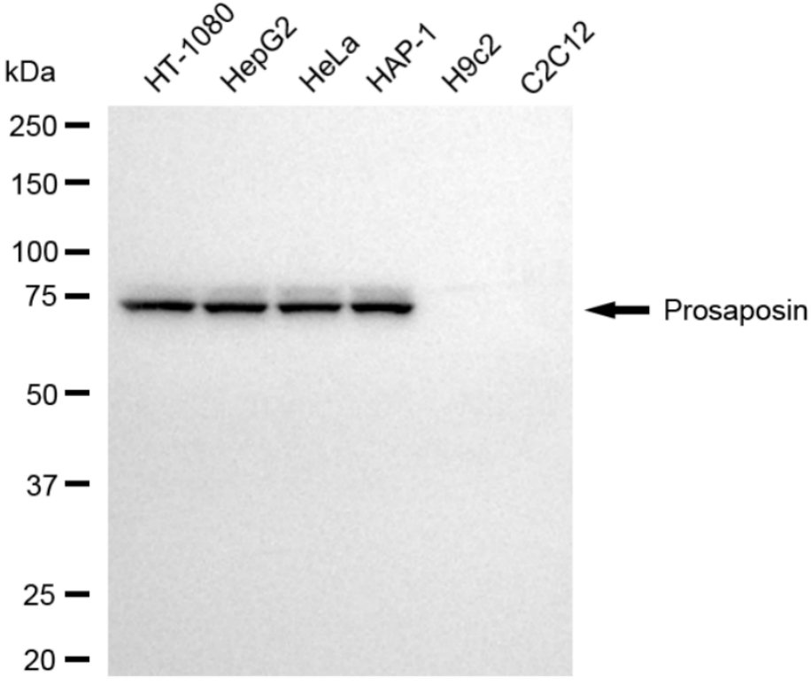
Western blotting analysis using prosaposin antibody. Total cell lysates (30 μg) from various cell lines were loaded and separated by SDS-PAGE. The blot was incubated with prosaposin antibody and HRP-conjugated goat anti-rabbit secondary antibody respectively.
PSAP Antibody (CSB-PA018836DA01HU)
驗證數據
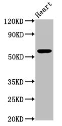
Western Blot
Positive WB detected in: Mouse heart tissue
All lanes: PSAP antibody at 3.3µg/ml
Secondary
Goat polyclonal to rabbit IgG at 1/50000 dilution
Predicted band size: 59 kDa
Observed band size: 59 kDa
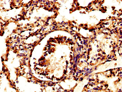
IHC image of CSB-PA018836DA01HU diluted at 1:500 and staining in paraffin-embedded human lung tissue performed on a Leica BondTM system. After dewaxing and hydration, antigen retrieval was mediated by high pressure in a citrate buffer (pH 6.0). Section was blocked with 10% normal goat serum 30min at RT. Then primary antibody (1% BSA) was incubated at 4°C overnight. The primary is detected by a biotinylated secondary antibody and visualized using an HRP conjugated SP system.
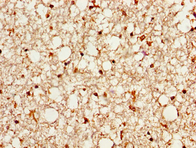
IHC image of CSB-PA018836DA01HU diluted at 1:500 and staining in paraffin-embedded human brain tissue performed on a Leica BondTM system. After dewaxing and hydration, antigen retrieval was mediated by high pressure in a citrate buffer (pH 6.0). Section was blocked with 10% normal goat serum 30min at RT. Then primary antibody (1% BSA) was incubated at 4°C overnight. The primary is detected by a biotinylated secondary antibody and visualized using an HRP conjugated SP system.
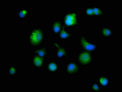
Immunofluorescence staining of MCF-7 cells with CSB-PA018836DA01HU at 1:166, counter-stained with DAPI. The cells were fixed in 4% formaldehyde, permeabilized using 0.2% Triton X-100 and blocked in 10% normal Goat Serum. The cells were then incubated with the antibody overnight at 4°C. The secondary antibody was Alexa Fluor 488-congugated AffiniPure Goat Anti-Rabbit IgG(H+L).
PSAP Antibodies
PSAP for Homo sapiens (Human)
| 產品貨號 | 產品名稱 | 種屬反應性 | 應用類型 |
|---|---|---|---|
| CSB-PA018836DC01HU | PSAP Antibody, FITC conjugated | Human | |
| CSB-PA018836DA01HU | PSAP Antibody | Human, Mouse | ELISA, WB, IHC, IF |
| CSB-PA018836GA01HU | PSAP Antibody | Human,Mouse,Rat | ELISA,WB,IHC |
| CSB-PA040013 | PSAP Antibody | Human | WB, IHC, ELISA |
| CSB-RA264216A0HU | PSAP Recombinant Monoclonal Antibody | Human | ELISA, WB, FC, ICC |
PSAP Proteins
PSAP Proteins for Homo sapiens (Human)
| 產品貨號 | 產品名稱 | 來源 |
|---|---|---|
| CSB-YP018836HU | Recombinant Human Prosaposin (PSAP), partial | Yeast |
| CSB-EP018836HU | Recombinant Human Proactivator polypeptide (PSAP), partial | E.coli |
| CSB-BP018836HU CSB-MP018836HU CSB-EP018836HU-B |
Recombinant Human Prosaposin (PSAP), partial | Baculovirus Mammalian cell In Vivo Biotinylation in E.coli |
PSAP ELISA Kit
PSAP ELISA Kit for Homo sapiens (Human)
| 產品貨號 | 產品名稱 | 樣本類型 | 靈敏度 |
|---|---|---|---|
| CSB-E12837h | human prosaposin(PSAP)Elisa kit | serum, plasma, tissue homogenates. | 0.041 ng/ml |